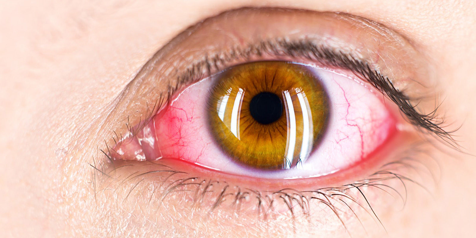
Diagnostics and treatment of retinal diseases
The retina is the neurological tissue of the eye and is the site of sight formation. Any disease that affects the retina can result in severe vision loss. Pigmentary retinopathy is a group of inherited retinal dystrophies that represent an important cause of vision loss in young people.
Dr. Marco Lombardo and his staff offer advice in the diagnosis and treatment of retinal diseases. At our Center the main exams are performed for a correct diagnosis of the patient and the choice of the most appropriate therapy.
Diabetic retinopathy
Diabetes mellitus consists of an abnormal concentration of glucose in the blood (values above 140 mg/dl). Diabetes is the leading cause of legal blindness in Italy and the western world.
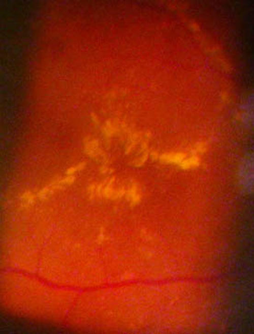
Diabetic macular edema
Controlling your blood sugar and undergoing periodic specialist visits is the best cure for preventing serious complications of diabetic retinopathy.It is well established that almost all people may have some signs of diabetic retinopathy after 20 years of diabetes onset. Late stages of diabetic retinopathy are accompanied by the most serious complications of the disease, such as secondary retinal detachment and neovascular glaucoma, both of which can lead to blindness.
The prevalence of diabetic retinopathy in the general population has dramatically increased over the last 40 years.
- Diagnosis and therapy of diabetic retinopathy
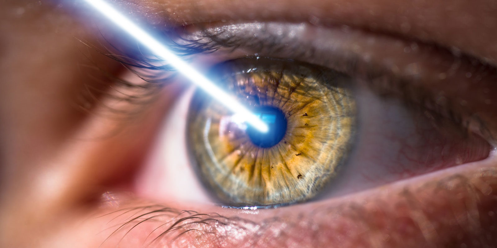
In the initial stages, diabetic retinopathy is usually asymptomatic. If the retinal disease worsens, visual acuity worsens caused by macular edema or episodes of vitreous hemorrhage. These complications require prompt intervention. An in-depth eye examination with retinal OCT is the most appropriate diagnostic pathway for an accurate assessment of diabetic retinopathy.
Treatment of diabetic macular edema consists in focal or grid laser treatment. In late stage of the disease, panretinal laser photocoagulation may be necessary. The aim of panretinal laser therapy is to reduce the risk of serious complications of diabetic retinopathy, such as hemovitreous (vitreous hemorrhage) and retinal detachment.
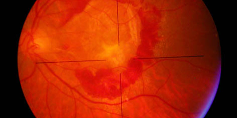
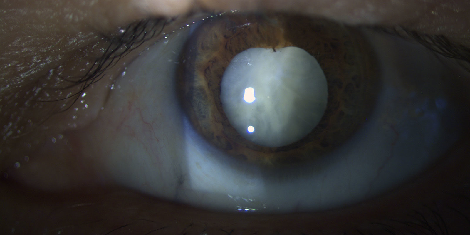
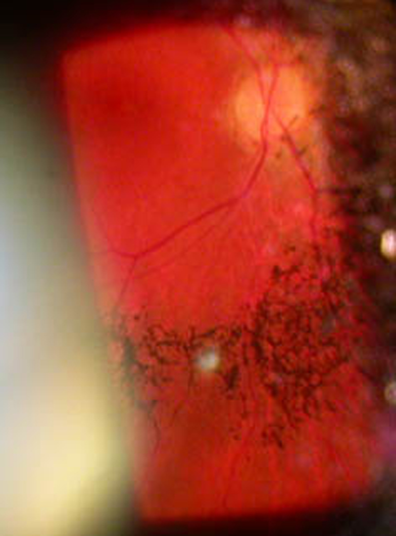
Retinal detachment
Retinal detachment consists in the separation of the retina from the retinal pigment epithelium. It may represent a serous emergency that needs immediate surgical care in order to avoid vision loss.
- Types of retinal detachment
The rhegmatogenous retinal detachment is the most frequent type of retinal detachment, and it is caused by one or more retina tears. The warning symptoms, may be represented by luminous flashes and floaters. The prevention of retinal detachment is very important and is based on periodic visits. The examination of the retina can detect the presence of retinal tears or degenerative areas in the retinal periphery.
Tractional retinal detachment is the most common cause of detachment in proliferative diabetic retinopathy.
In the exudative retinal detachment, the subretinal fluid creates the retinal detachment. The causes can include intraocular tumors (choroidal melanoma, metastasis), inflammatory diseases (chorioretinitis), congenital anomalies (Coats disease) or degenerative macular diseases. An ocular ultrasound is always required to proper preoperative management of retinal detachment. - Retinal detachment surgery
There are various surgical procedures, including scleral buckling and vitrectomy to fix a retinal detachment. The scleral buckling is still today the most used surgical procedure; vitrectomy is performed to remove vitreous tractions.
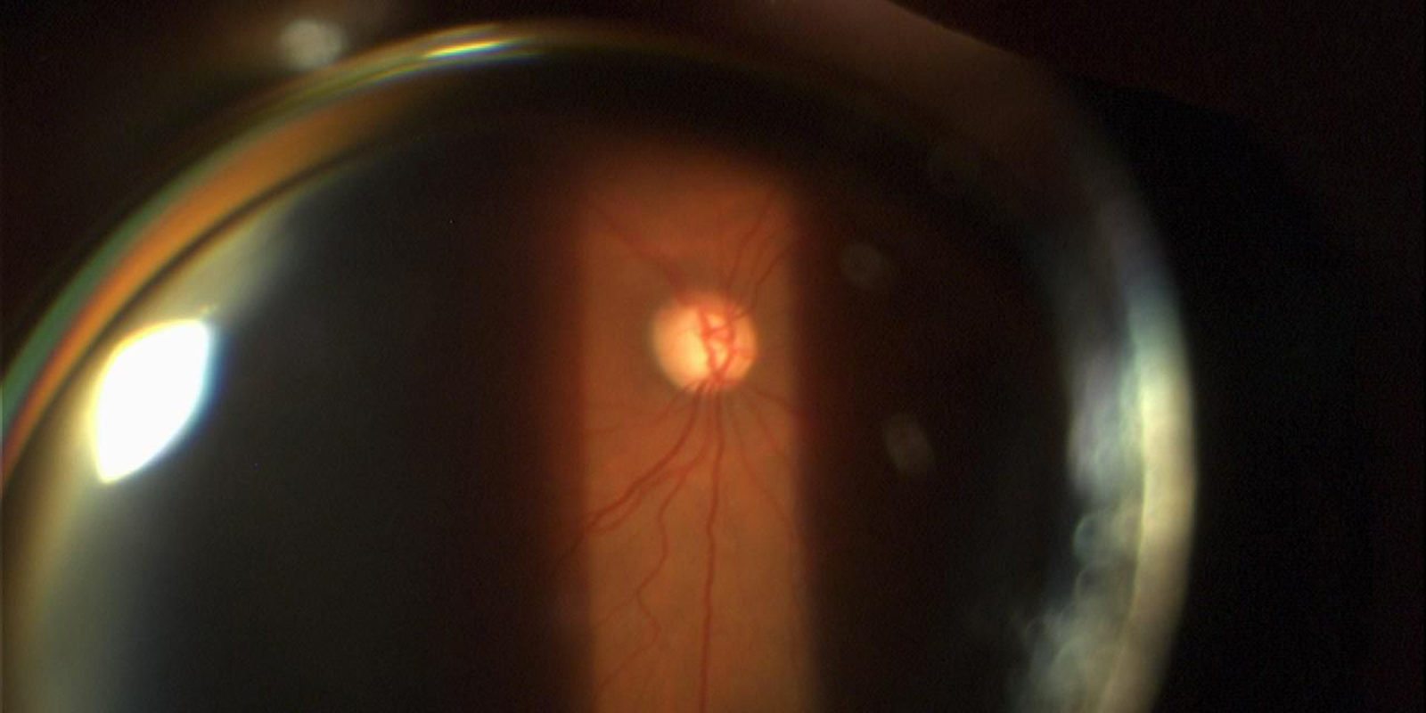
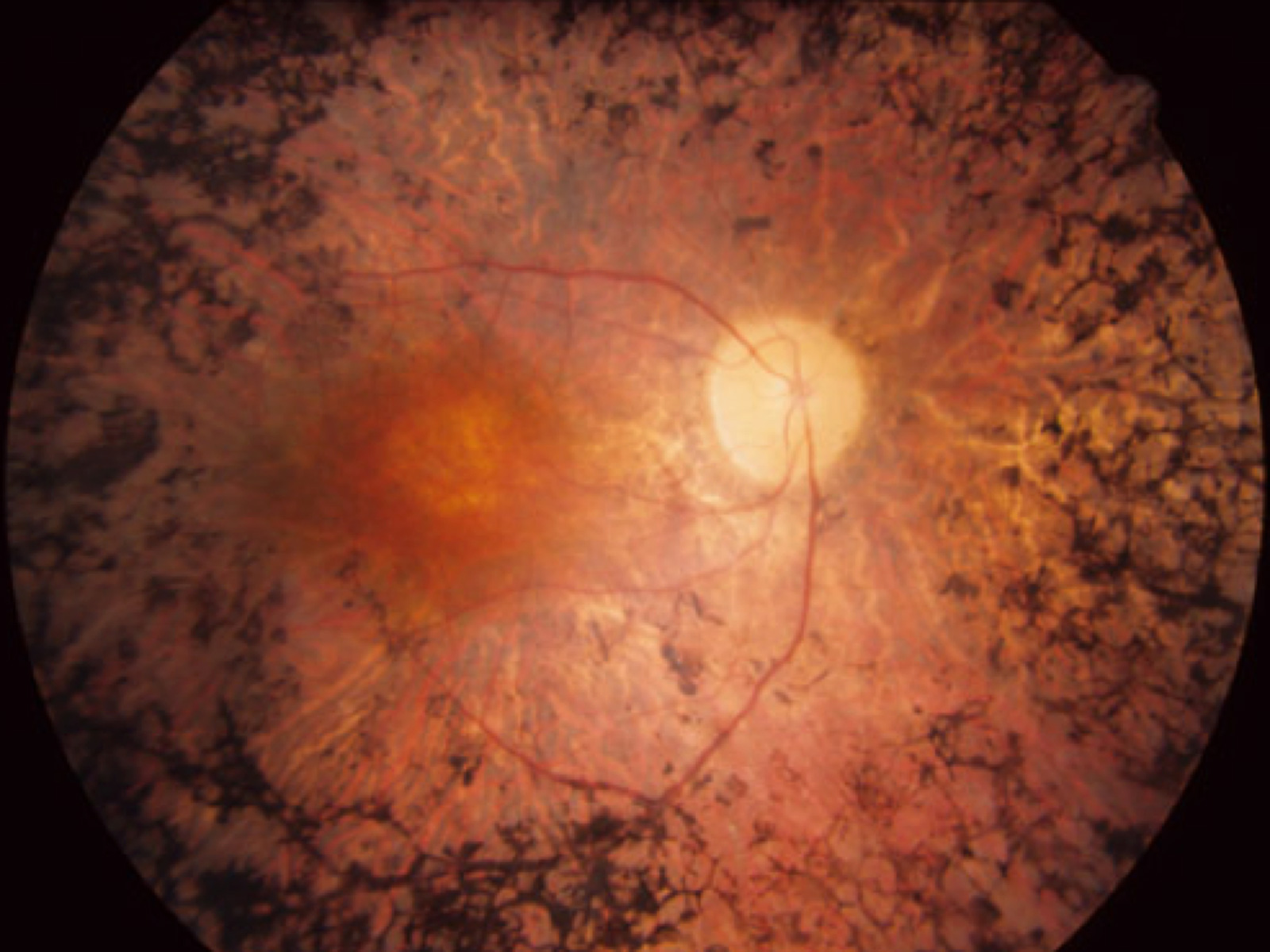
Pigmentary retinopathy
The term pigmentary retinopathy contains a large group of hereditary retinal degenerations in which the retinal photoreceptors and the retinal pigment epithelium are primarily affected.The age of onset of symptoms may vary and is associated with the type of genetic transmission.
- Diagnosis of pigmentary retinopathy
Pigmentary retinopathy is one of the main causes of visual impairment in the young and adult age in the western world. The initial symptom may vary in accordance with the form of disease; visual acuity is usually preserved up to the most advanced stages of the disease.
The diagnosis of pigment retinopathy is established when the following conditions are present:- bilateral involvement;
- loss of peripheral vision;
- progressive loss of photoreceptor function determined by electroretinogram(ERG).
Pigmentary retinopathy
- Pigmentary retinopathy therapy
There are currently no therapies able to halt or regenerate the atrophic retinal areas. Patients today can benefit from the use of optical aids: polarized lenses, telescopic systems, wide angle optical groups (for expand the visual field). Diet supplementation with vitamin A, cyanosides, omega-3 fatty acids, lutein, curcumin can improve the metabolic conditions of the degenerated retinal areas but there is no evidence that their use can stop disease progression. Gene therapy, retinal cell transplantation and retinal prosthesis implantation represent the next golden standard for the treatment of pigmentary retinopathy.